
Spinal Stenosis Treatment
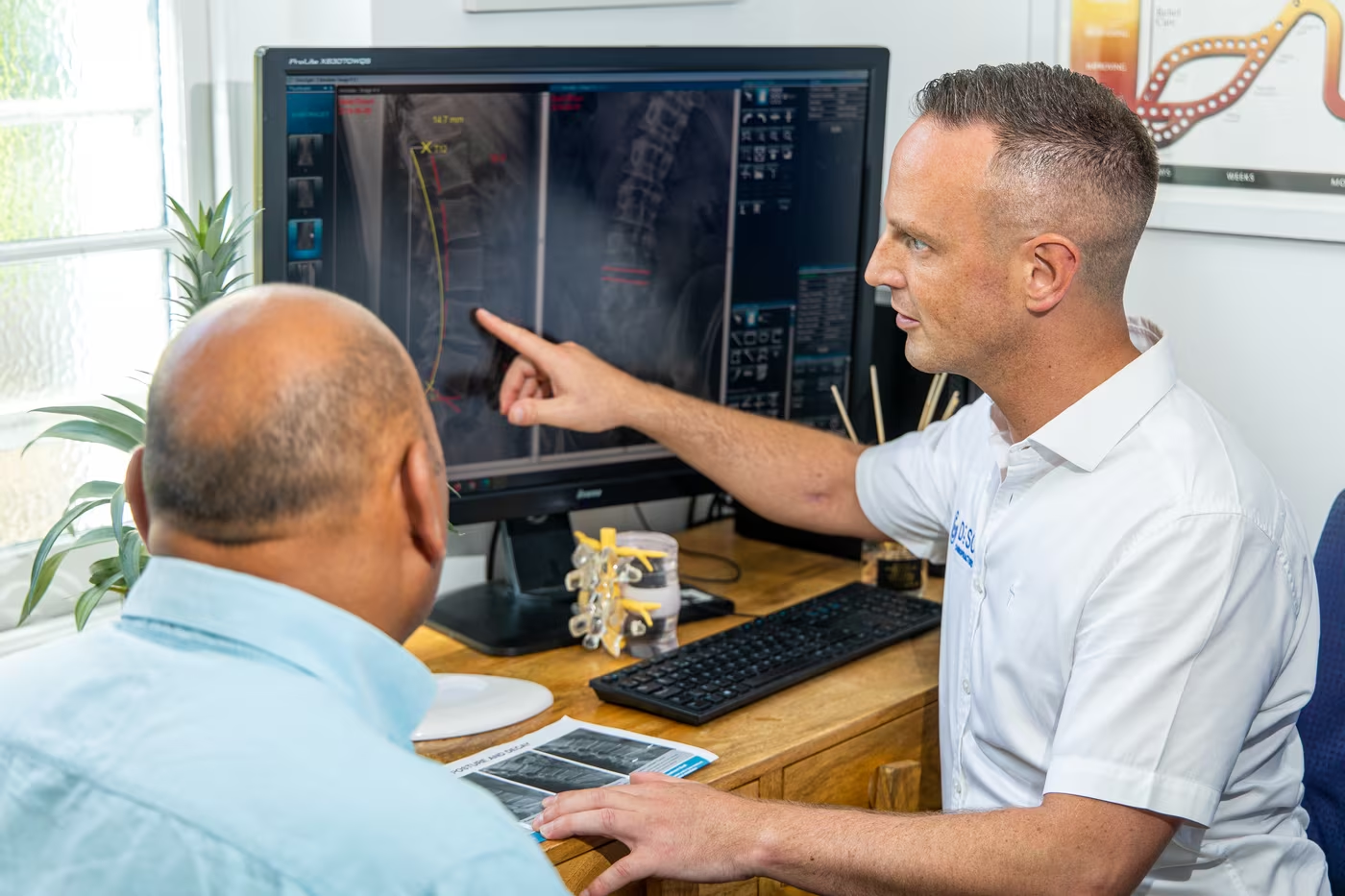
Most people meet this diagnosis as a sentence on a scan report. Spinal stenosis. Canal narrowing. Foraminal narrowing. Moderate to severe at L4-5.
The picture that forms is almost always the same. Nerves being squeezed, a spine closing in on itself, a process that can only go one way. Surgery will be needed eventually, but it can take years of suffering before it’s considered serious enough to operate.
Here is what does not fit that picture. How narrow the canal looks on a scan does not reliably predict how much pain you will have, or how far you will be able to walk. Some people have considerable narrowing with surprisingly little trouble, while others with more moderate changes are significantly limited.
Clinically, our experience matches this duality: I have seen severe-looking scans that get miraculous relief within days, and I’ve had cases that seem to struggle despite X-rays looking reasonable.
What is spinal stenosis?
Stenosis means narrowing. The word describes a space rather than a disease.
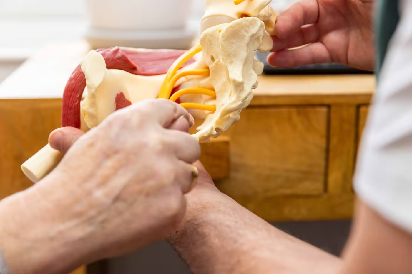
Discs lose height and settle, or sag and bulge. The facet joints at the back of the spine, which take more load as disc height reduces, enlarge and become arthritic. The ligament running along the back of the canal thickens and buckles inwards. Bone spurs form at the joint edge. Sometimes one vertebra slips slightly forward on the one below.
None of these alone is spinal stenosis. Stenosis is what happens when enough of them accumulate in the same place that the space starts to matter.
That is why describing stenosis as just advanced disc degeneration is misleading. Without including the implications of the narrowing and its effects on the nerves that transit adjacent to the structures.
“Spinal stenosis is the most common cause of leg pain and disability in adults over the age of 50, with approximately 2 million people affected each year.”
American Academy of Orthopaedic Surgeons
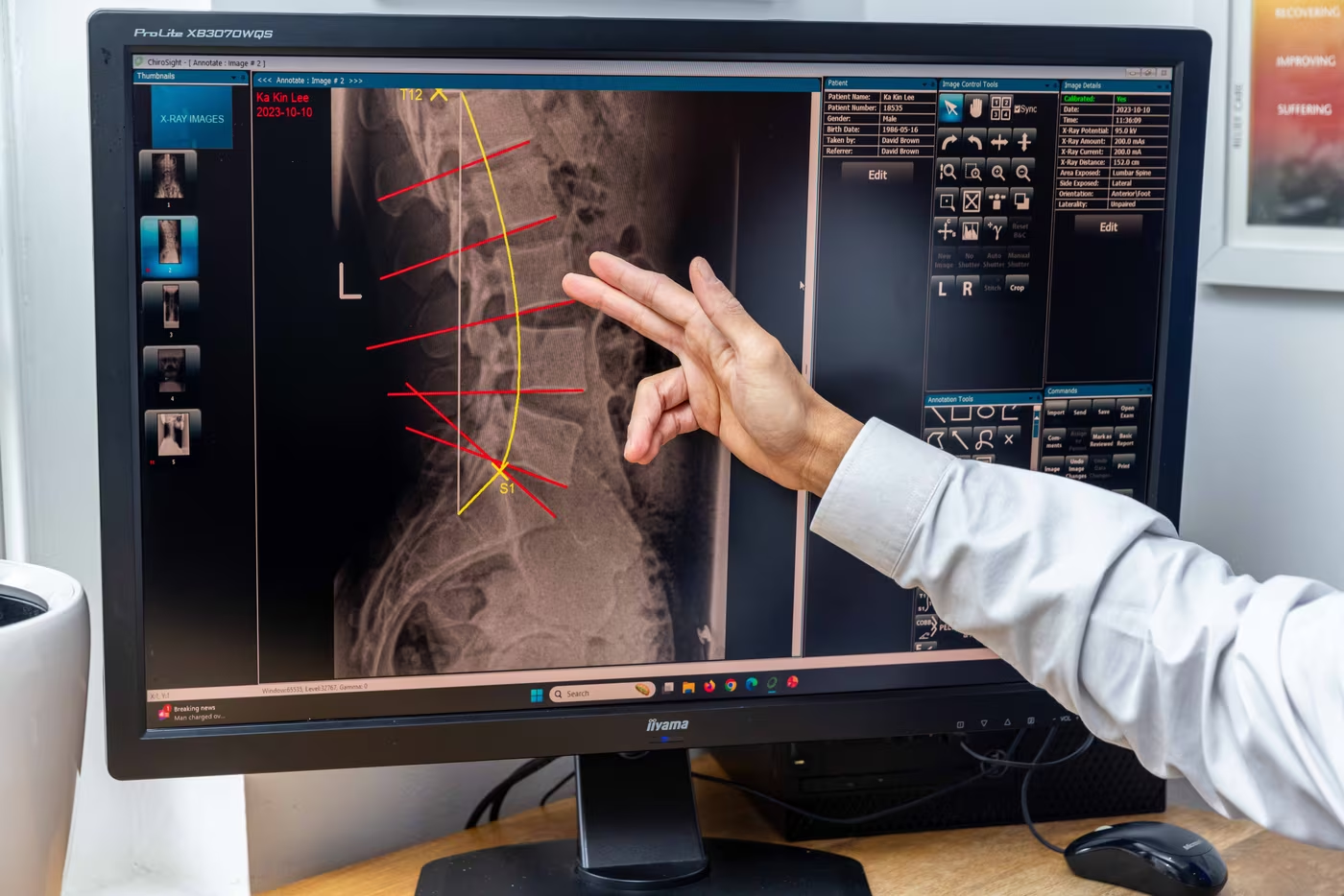
Relevance: Does the Spinal stenosis on your scan explain your symptoms?
This is the question our whole assessment exists to answer, and it gets less attention than it deserves.
Radiological stenosis is common. Pooled across studies, around one in ten people with no symptoms at all have imaging-defined stenosis, and the figure climbs steeply with age. A report describing narrowing in a 72-year-old is closer to an expected finding than to an explanation.
More striking is what happens when severity is compared against how people actually are. A grading system based on how crowded the nerve roots look on MRI found no correlation between the grade and the patient’s level of disability. The relationship is not merely weak. It sometimes runs backwards.
That does not make the narrowing irrelevant. Severity carries some weight, but the scan is one piece of information, not the answer. What makes a narrowing clinically meaningful is whether the symptoms behave as if they are due to compressed neural tissue, whether the examination agrees, and whether the level involved matches the location of the symptoms.
What spinal stenosis feels like, and why walking gives it away
The pattern has a name, neurogenic claudication, and it is the most useful thing on this page for working out whether stenosis is your problem.
It usually starts in the legs rather than the back. People describe heaviness, aching, burning, pins and needles, numbness or a sense that the legs are simply not obeying. It comes on with standing and walking, builds the longer you continue, and eases when you sit down or bend forward.
Back pain may be prominent or barely present. Walking distance is often telling and can vary, better on some days and some routes than others, which usually makes sense once you look at the terrain.
These features carry real diagnostic weight; however, a review also found that the syndrome is better at ruling out stenosis than at ruling it in. If the pattern is absent, stenosis becomes considerably less likely. If it is present, it makes stenosis likely, though it does not confirm it.
The shopping trolley sign is one of the most characteristic signs of lumbar spinal stenosis, particularly in older patients. Someone may struggle to walk upright but find they can go considerably farther when leaning forward over a shopping trolley, a walking frame, or even a garden mower. Cycling can produce the same effect: a person who can only walk a short distance may be able to cycle surprisingly comfortably.
There is a mechanical reason for this. Bending forward increases the available space within the lumbar spinal canal and around the exiting nerves, while extending backwards reduces it. In a spine where that space is already limited by enlarged facet joints, thickened or buckled ligaments, disc changes and other degenerative narrowing, even a relatively small change in posture can matter.
Is spinal stenosis more than just a crush?
Historically, the mechanism of spinal stenosis was explained mainly as a space problem. Standing and walking, both upright postures, tend to place the lumbar spine under compression, reducing the relative space within the spinal canal and around the nerve roots. Sitting, bending forward or leaning over a shopping trolley moves the spine into flexion and increases that space again. This remains an important part of the explanation.
Current thinking is more nuanced. Spinal stenosis appears to behave as a dynamic condition rather than simply a fixed narrowing visible on an MRI.
There is also good reason to think that compression is not the whole story. The alternative theory is a pattern called neurogenic claudication: the legs become heavy, painful, numb, or weak with prolonged standing and walking, even over short distances, then improve with sitting or bending forward.
The nerve roots are already short of space. Upright loading and repeated movement can increase local pressure, impairing blood flow to the nerves, effectively starving them. This may temporarily limit their ability to function normally, which helps explain why symptoms worsen with walking and settle with rest, when pressure is reduced and circulation can recover.
The precise contribution of each mechanism is still being debated amongst researchers. The important change in our understanding is that lumbar stenosis is no longer best thought of simply as a pipe that has become too narrow. It is the interaction between the available space, posture, loading, and the nerves’ ability to continue functioning within that space that helps explain why someone may feel relatively comfortable sitting down yet become increasingly symptomatic the further they walk, unfortunately compounding behaviours that promote less movement.
What else can cause leg symptoms when you walk
Leg symptoms brought on by walking are not automatically indicative of spinal stenosis, and misdiagnosis can waste months of effort.
Vascular claudication comes from the circulation to the leg rather than the nerve. It sits in the calf, comes on at a fairly predictable distance regardless of posture, and eases when you stand still rather than needing you to sit or bend. Pulses may be reduced, and leaning on a trolley makes no difference. This matters because it needs a different speciality.
Peripheral neuropathy, often related to diabetes although it can be brought on by other factors, tends to be symmetrical, glove-and-stocking in distribution, and present whether you are moving or not.
These overlap more than textbooks suggest, and it is entirely possible to have two of them at once. That is a large part of why the examination matters.

Can’t surgery just fix my spinal stenosis?
Surgery for spinal stenosis can do something conservative treatment cannot. It can physically create more space around the neural structures. In extreme cases, that is a real advantage, and it should not be talked down.
Surgical opinion becomes increasingly appropriate when function is substantially limited, walking reserve is continuing to fall, there is progressive neurological deficit, properly delivered conservative care has not produced enough improvement, and the imaging genuinely corresponds with the clinical picture.
But spinal stenosis predominantly affects an older population, often with other health problems. So, statistically, not having surgery does not necessarily mean someone does not need or want it. For some patients, age, frailty, other medical conditions, or the complexity of the proposed operation makes the risk unacceptable. Others decide that surgery is simply not a risk they are prepared to take.
In either situation, conservative treatment is not pretending that the narrowing can be removed. The aim is to preserve and, where possible, increase the functional reserve available in the remaining space. In undertaking a course of rehabilitation, even if it fails, it will strengthen the back and legs, potentially allowing for a better surgical outcome and a better ability to heal up after the surgery
Additionally, the choice is not simply whether to operate, but what operation is appropriate. A laminectomy or decompression removes bone and ligament that are narrowing the canal, creating more room for the nerves. Fusion adds screws, rods, and bone graft to stabilise the spine and is usually considered when there is significant instability, a vertebral slip, deformity, or mechanical back pain that decompression alone is unlikely to address.
Fusion is a larger operation, with greater blood loss, longer recovery and more potential complications. It is necessary if removing the required amount of bone would destabilise the area, or if slippage is already occurring.
The failure rate of surgery is an obvious concern, although it is not a single fixed number. Outcomes depend on the operation performed, the underlying diagnosis, the patient’s general health, and the severity and duration of symptoms.
Persistent pain, recurrent narrowing, adjacent-segment disease, non-union after fusion, infection, nerve injury and the need for further surgery are all possible outcomes that should be considered.
A technically successful operation can therefore still produce a disappointing result if the main source of pain was not the compressed nerve, if back pain predominated, or if the patient’s functional limitations were caused partly by other conditions.
This is why the decision should not be based on the MRI report alone, or on a quoted success rate detached from the individual circumstances. The relevant question is whether the proposed operation addresses the symptoms that matter most, whether the expected benefit is large enough to justify the risks, and what the realistic alternatives are if the result is incomplete or symptoms later return.
What else could be causing leg symptoms when you walk?
Leg symptoms brought on by walking are not automatically indicative of spinal stenosis, and misdiagnosis can waste months of effort.
Vascular claudication comes from the circulation to the leg rather than the nerve. It sits in the calf, comes on at a fairly predictable distance regardless of posture, and eases when you stand still rather than needing you to sit or bend. Pulses may be reduced, and leaning on a trolley makes no difference. This matters because it needs a different speciality.
Peripheral neuropathy, often related to diabetes although it can be brought on by other factors, tends to be symmetrical, glove-and-stocking in distribution, and present whether you are moving or not.
These overlap more than textbooks suggest, and it is entirely possible to have two of them at once. That is a large part of why the examination matters.
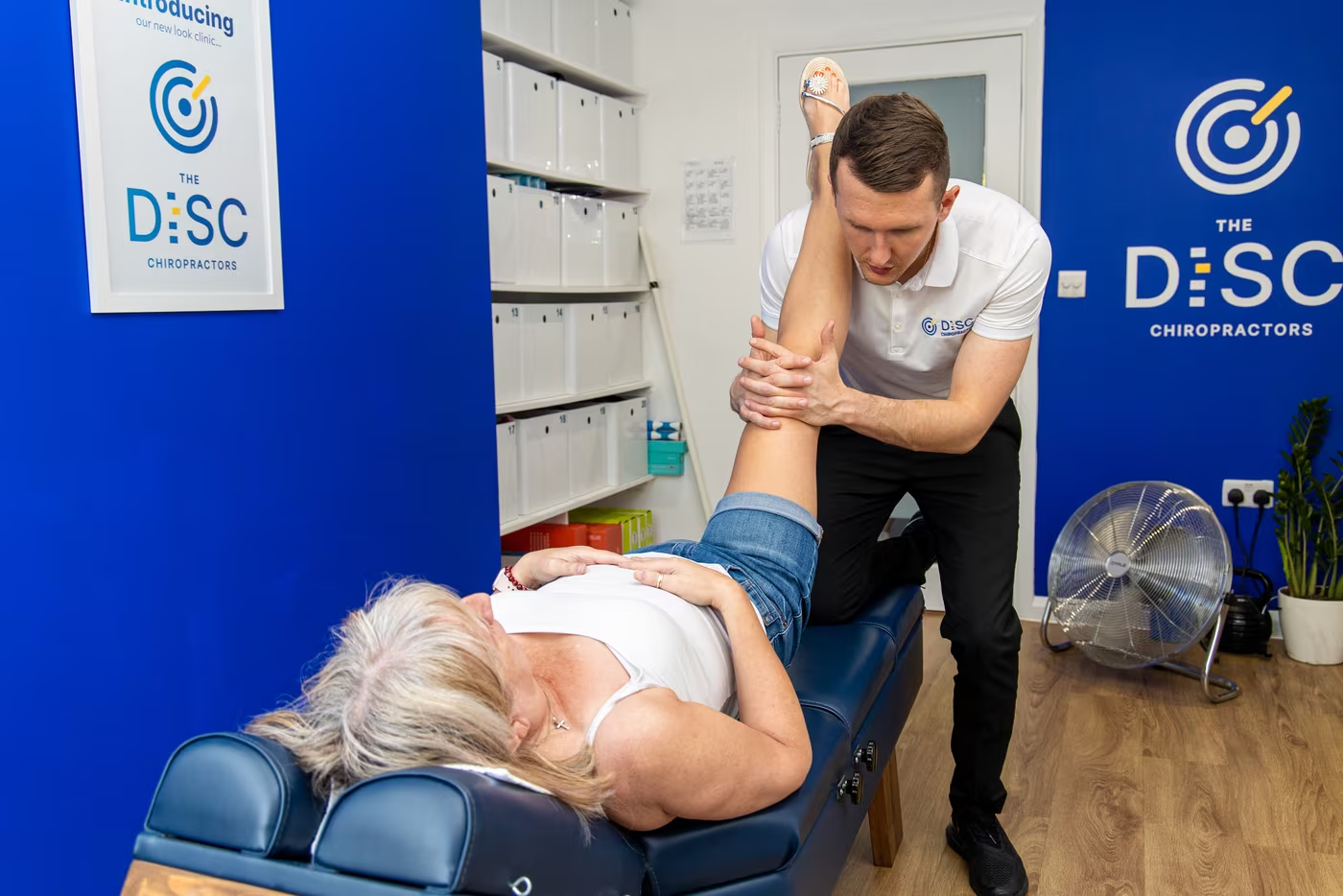
What should you avoid with spinal stenosis?
Almost certainly fewer things than you have been told.
Most people arrive with fears assembled over several years, and the cumulative effect is a spine that does progressively less and tolerates progressively less. Rest is not a treatment for spinal stenosis. Three situations get muddled together and separating them is most of the answer.
During a bad patch, temporary modification is sensible. Breaking up long periods of standing, using a trolley or poles, choosing flatter routes, sitting before the symptoms force you to. This is temporary and should be self-limiting.
Rest does not mean stopping moving; it just means avoiding known stressors.
Where a specific position or load reliably provokes symptoms, that is information rather than a prohibition. Sustained standing and walking in extension are the usual culprits, and both can often be modified rather than abandoned.
Everything else is worth keeping.
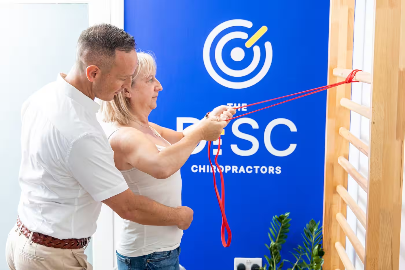
What Rehabilitation exercises are appropriate for Spinal Stenosis
Rehabilitation for spinal stenosis should have a very practical goal: help you stand and walk further before your symptoms make you stop, not to reverse bony changes, as that’s impossible.
One of the better-known rehabilitation approaches was developed by spinal stenosis researcher Carlo Ammendolia. His programme combines flexion-biased exercises, strengthening, soft-tissue work, stretching, cycling, walking and strategies designed to reduce excessive lumbar extension.
Progress is measured partly by how far the patient can walk, rather than simply by whether their pain score has changed.
The answer isn’t simply to avoid anything that triggers symptoms. Nor do we think everyone with stenosis needs the same sheet of pelvic tilts and stretch exercises.
Rehabilitation should match what that individual has lost. That might mean improving leg and trunk strength, using cycling or other flexion-tolerant exercise to rebuild cardiovascular fitness, improving balance, changing positions that unnecessarily close the available space, and gradually increasing walking exposure.
Walking itself eventually needs to be part of the rehabilitation. If someone currently develops symptoms after 200 metres, repeatedly forcing them to walk until their legs become unbearable is unlikely to be a clever training strategy. But permanently restricting them to 200 metres also guarantees their walking capacity is never challenged.
We therefore use walking reserve as both part of the problem and part of the measurement. Can you walk further before symptoms begin? Can you continue for longer once they start? Do they settle more quickly when you stop? Can you repeat the activity after a shorter recovery?
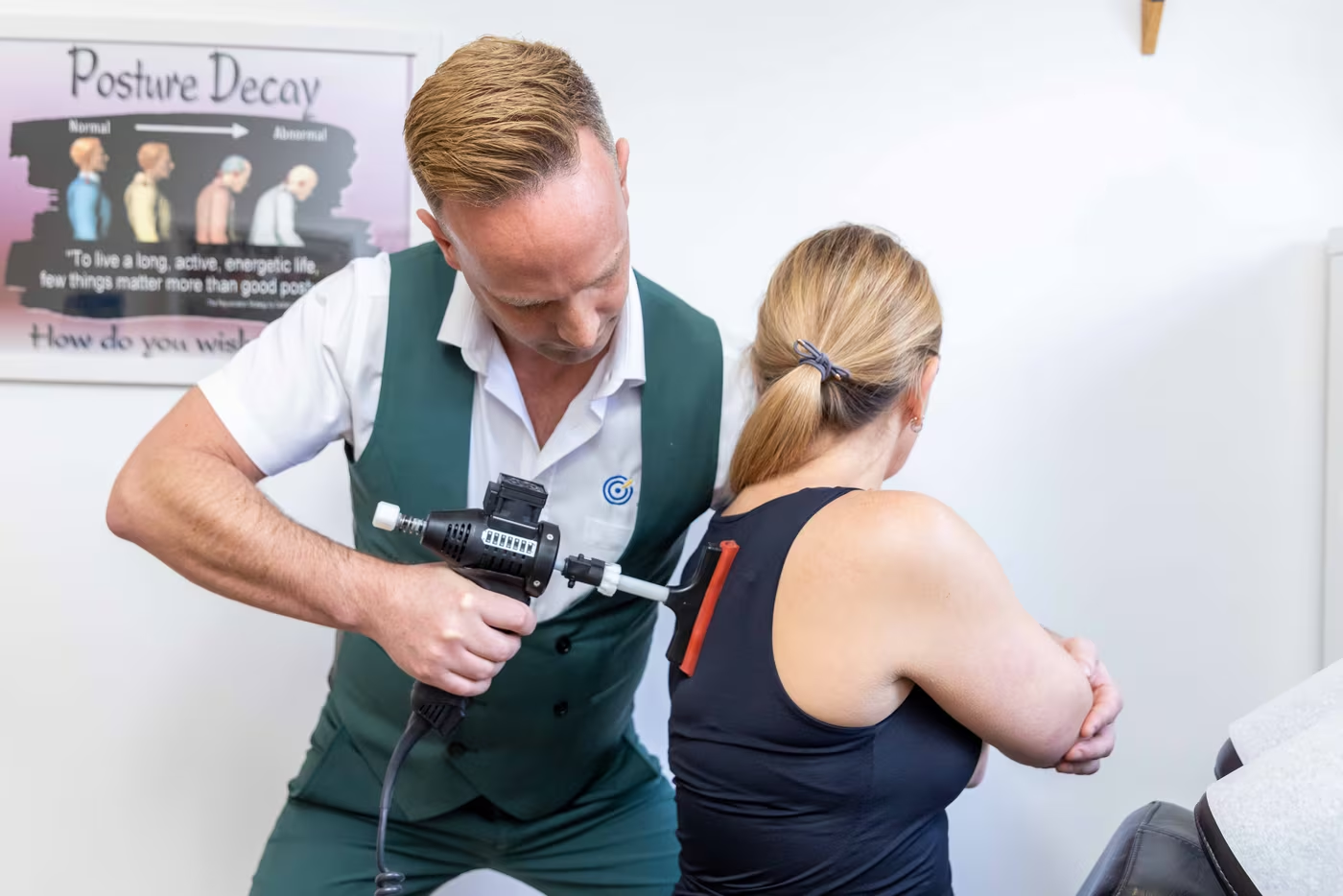
Can a Chiropractor Help With Spinal Stenosis?
Yes, but lumbar spinal stenosis requires a more specialised approach than routine chiropractic care. It is not the same as treating a disc or joint problem, and traditional chiropractic treatment alone will rarely be enough to meaningfully change an advanced stenosis.
The stenosis itself cannot be manipulated away.
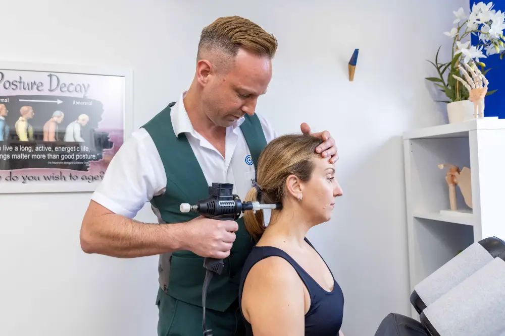
Where conservative treatment is appropriate, a specialist’s knowledge of stenosis rehabilitation, using specific drop table adjustments, flexion-distraction tables (relatively common), or advanced spinal decompression therapy (relatively rare), which, whilst not technically a chiropractic technique, is insanely valuable and gaining momentum in these cases.
The focus is on the factors that remain changeable around the narrowing. That may include restricted movement above or below the affected level, stiff hips, difficulty returning to a comfortable upright posture, muscle guarding, loss of leg and trunk strength, and the general deconditioning that can develop after months or years of reduced walking.
Not every patient has all of these problems. The examination remains crucial to determine what is actually present, how much of it contributes to the symptoms, and whether chiropractic treatment is appropriate at all.
Much of the knowledge and experience we have gained in treating spinal stenosis has been in clinic, adapting our unique disc injury protocol to fit more degenerative cases. Likewise, manual therapy/chiropractic has generally been studied in spinal stenosis as one component of a broader programme alongside exercise and progressive walking, rather than as a standalone solution. The studies reporting meaningful improvements in walking have used that combined approach; this is a key component of our stenosis protocol.
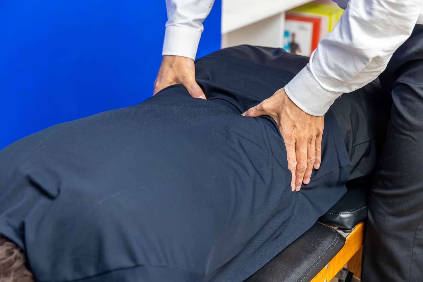
Can Chiropractic Make Spinal Stenosis Worse?
Potentially, but there isn’t a single medical or chiropractic technique that is 100% safe. If the wrong treatment is applied to the wrong patient. That is precisely why spinal stenosis should not be treated as routine mechanical back pain.
Where treatment is appropriate, technique should reflect the patient’s age, anatomy, and irritability. An older spine with advanced degenerative change may require a very different approach from an otherwise healthy lower back.
Any pain after manual therapy is usually very short-lived and resolves within 24 hours. There is no obvious reason why chiropractic treatment for spinal stenosis carries a significantly higher risk than other cases, if the techniques involved are moderated for the patient’s age, as they always should be regardless of the diagnosis.
Why Is Spinal Decompression Therapy Such a Good Fit for Spinal Stenosis?
Spinal decompression therapy is one of the treatments we find particularly useful for managing lumbar spinal stenosis because its mechanism aligns well with how stenosis behaves.
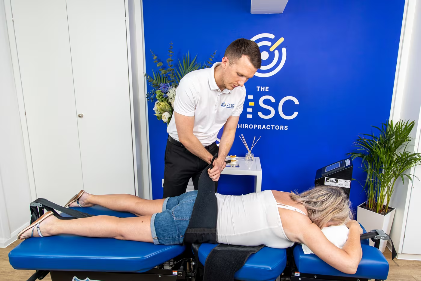
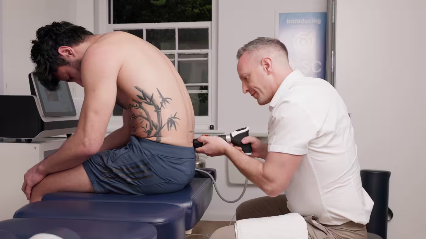
Stenosis is characteristically aggravated by loading. Standing and walking reduce the available space around already-compromised nerves, while sitting, flexing forward, and unloading the spine often bring relief. Spinal decompression takes that principle considerably further by applying controlled, targeted unloading to the affected region while the patient lies comfortably.
Nobody is claiming that decompression physically removes the stenosis. But its axial traction is an obvious benefit to opening up the spinal segments as much as possible. Decompression gives us another way to work with the spine while we progressively improve movement, strength and walking tolerance around it.
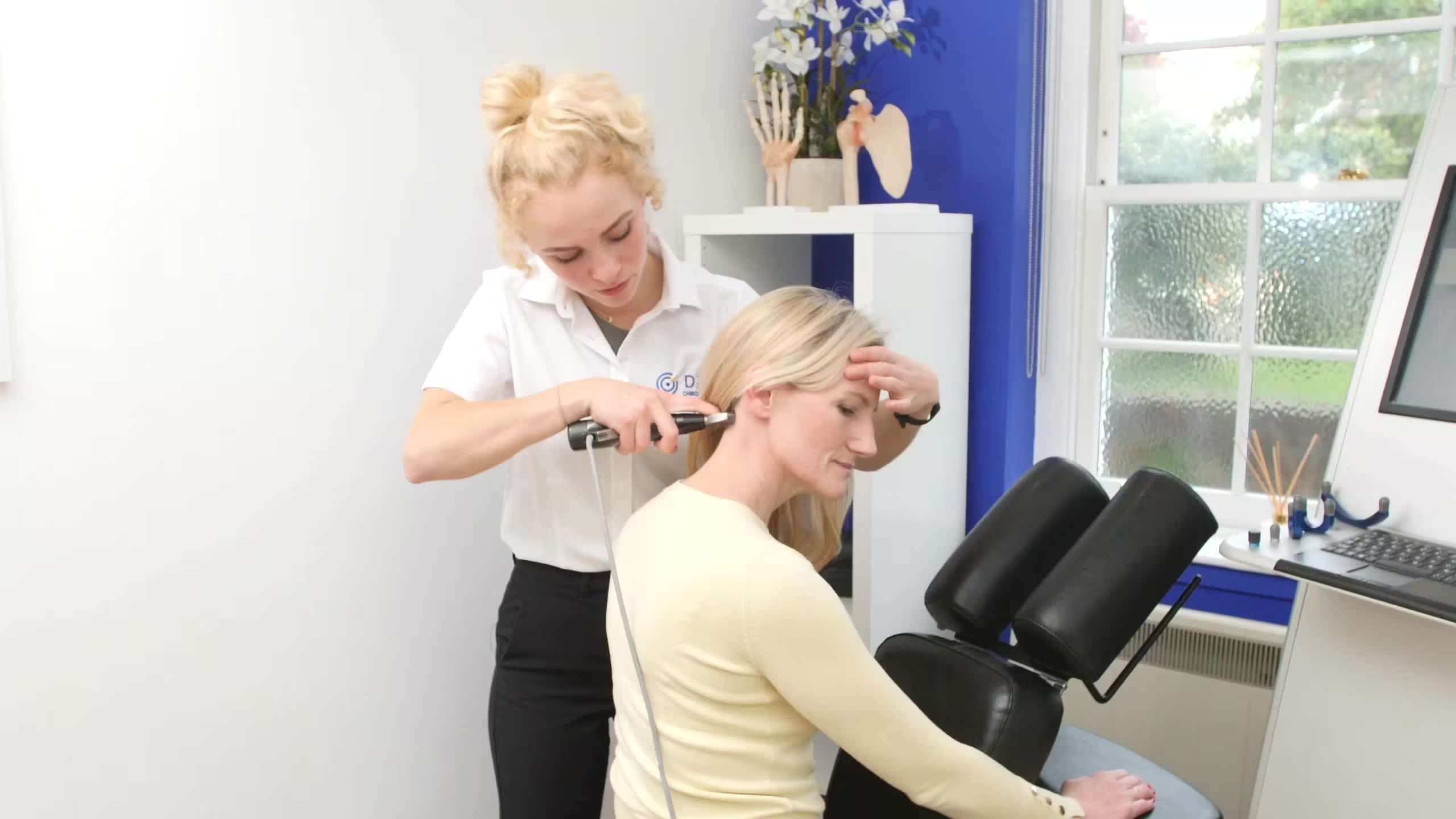
We rarely view spinal decompression as a treatment in isolation. Within our stenosis protocol, it can be combined with appropriate chiropractic techniques, rehabilitation, and progressive walking, with each component serving a different role.
What about stenosis in the neck?
Narrowing in the neck can be a different proposition.
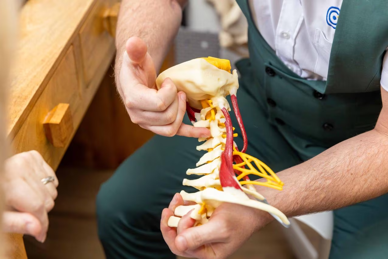
In the lower back, it affects nerve roots. In the neck, it can affect the spinal cord itself, which changes what needs to be monitored. Cord compression visible on imaging is relatively common and mostly asymptomatic, present in about a quarter of people without relevant symptoms and in over a third of those over 60. Only a small proportion develop problems from it.
Where the cord is genuinely affected, the condition is known as degenerative cervical myelopathy, and its early signs are quiet. The hands usually give it away first. Dropping things, fumbling with buttons, deteriorating handwriting, difficulty with cutlery or coins. Around 85 per cent of people with the condition have some hand symptom. Alongside that, changes in balance and walking, unsteadiness in the dark, or symptoms in both arms or both legs.
These are not features of ordinary neck pain. If they are developing or worsening, that is a reason for prompt medical assessment rather than a course of treatment, and it is something we screen for at every neck assessment, including a simple grip-and-release test that takes ten seconds.
Guidance on when the neck needs surgery is clearer than for the lower back: severe myelopathy warrants surgery.
Mild symptoms should be handled with manual therapy and rehabilitation; moderate symptoms can be managed similarly, with the knowledge that if symptoms do not improve, referral back to the medics is advised.
Will my spinal stenosis get worse?
The honest answer is that the narrowing itself usually does, but your symptoms and function do not necessarily follow the same path.
So, care and rehabilitation are not aimed at making the scan look normal. It is designed to help you function better within the remaining space. This is both recommended and achievable for those willing to put in the effort.
That may involve improving leg and trunk strength, gradually building walking tolerance, working on balance and confidence, modifying activities that repeatedly aggravate symptoms, and finding positions or movement strategies that allow you to do more with less discomfort. The programme should be adjusted based on your response, not solely on the severity of the MRI findings.
Long-term studies of people managed without surgery show a much less predictable course than imaging might suggest. Some improve, some remain remarkably stable for years, and others progressively lose walking tolerance and function.
Rehabilitation cannot guarantee which path an individual will take, but it can often improve symptoms, walking tolerance and confidence, giving patients a fighting chance.
With spinal stenosis, progress is better judged by what you are gaining or losing in the real world than by whether the adjective on the MRI changes. It is therefore generally accepted that the distance you can walk is the yardstick by which spinal stenosis is graded.
What to do next?
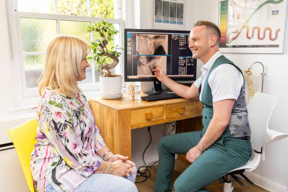
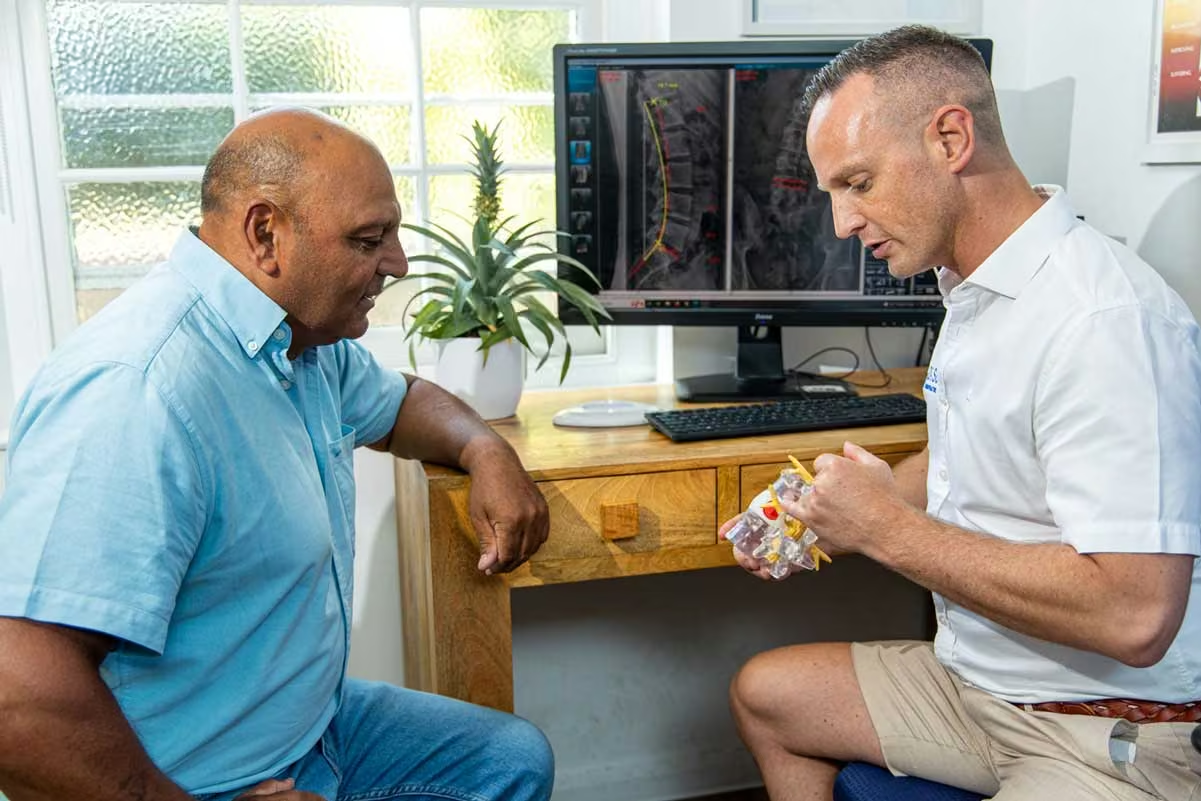
If an X-ray or scan has told you that you have spinal stenosis, the useful next step is an assessment answering four things. How much room there actually is. Whether that narrowing is causing your symptoms or coexisting with them. How much walking and standing reserve you have. And whether that reserve is holding, drifting or falling.
Those answers decide everything that follows, including whether it’s appropriate to take the case on as a patient. Some people need an explanation and better information about how to walk. Some need a short course of care. Some need a staged programme to rebuild tolerance. A few need a surgical opinion sooner than they have been led to believe. Knowing which you are is worth more than any single treatment.